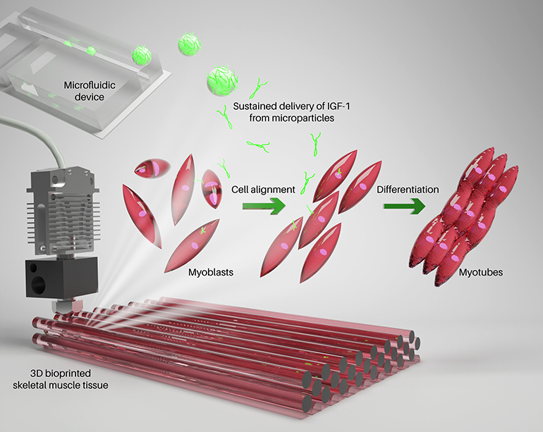

Researchers on the Terasaki Institute in Los Angeles have developed a brand new technique to create 3D printed muscle constructs with enhanced muscle cell alignment and maturation. The method includes creating microparticles loaded with insulin-like development issue (IGF) utilizing a microfluidic platform. Then, these particles are included in a bioink that additionally incorporates myoblast cells and a gelatin-based hydrogel. As soon as 3D printed, the ensuing constructs present enhanced cell development, elongation, and alignment, and in some instances even started to spontaneously contract after a ten day incubation. The Terasaki researchers hope that their innovation will assist pave the best way for absolutely useful, lab-created muscle transplants for human sufferers.
Skeletal muscle is clearly essential for motion and primary exercise. If such muscle turns into injured or must be eliminated due to harm or illness, then a affected person’s high quality of life can change considerably as their potential to maneuver and carry out every day actions is affected. Furthermore, different intently related tissues, comparable to lymph or blood vessels, may additionally be affected, resulting in further problems. At current, the primary remedy choice is to take away wholesome muscle from elsewhere within the physique and transplant it to the area the place it’s required.
Nevertheless, this isn’t supreme. Not solely is that this technique extremely invasive, damaging wholesome tissue to restore an harm elsewhere, however it could possibly have blended outcomes, with points comparable to incomplete innervation affecting the transplant efficiency and limiting the exercise of the transplanted muscle. These points have prompted scientists to aim to create lab-grown alternate options utilizing biomaterials.
3D bioprinting represents a really helpful method on this context, permitting researchers to print constructs in numerous sizes and styles very quickly. These researchers used this method, however enhanced it with the considered inclusion of slow-release development elements to affect cell exercise inside the assemble.
They included microparticles within the bioink that launch IGF slowly inside the assemble over a interval of days, serving to to steer the included myoblasts cells in direction of a skeletal muscle phenotype. Thus far, the tactic seems to assist in encouraging the cells to elongate and align, similar to the true factor, and a few constructs even demonstrated muscle contractions.
“The sustained launch of IGF-1 facilitates the maturation and alignment of muscle cells, which is a vital step in muscle tissue restore and regeneration,” mentioned Ali Khademhosseini, a researcher concerned within the examine. “There may be nice potential for utilizing this technique for the therapeutic creation of useful, contractile muscle tissue.”
Research in journal Macromolecular Bioscience: Enhanced Maturation of 3D Bioprinted Skeletal Muscle Tissue Constructs Encapsulating Soluble Factor‐Releasing Microparticles
By way of: Terasaki Institute
Trending Merchandise


![[2025 Upgrade] Aotedor 30 Miles Long Travel Range, Electric Wheelchair for Adults Power Wheelchairs Lightweight Foldable All Terrain Motorized Wheelchair for Seniors Compact Portable Airline Approved](https://m.media-amazon.com/images/I/51vZJPDMrOL._SS300_.jpg)
